Three Ways OptiLight Is Changing Dry Eye Management in Our Practice


The ethos of our practice is to help people be the best version of themselves, by addressing pathology and rejuvenating and restoring appearance. One focus area and subject of our current research is ocular surface and dry eye disease, for which we now offer a whole new level of care with OptiLight, which forms a core part of our OptimEyes™ program.
In London, where heredity makes many people likely to suffer with some degree of rosacea, blepharitis, and associated dry eye disease, we are ideally located to research the gold standards treating dry eye.
Benefits of OptiLight for Dry Eye Treatment
In our effort to provide quality, evidence-based care for our patients, we were motivated to get OptiLight because the technology is supported by over 40 clinical studies, differentiating it from other treatment options. Now, for the first time, we can manage dry eye disease effectively with OptiLight, not just by controlling symptoms, but by targeting the most common cause, meibomian gland dysfunction. OptiLight is already changing our practice in three important ways.
Superior engineering translates to better treatment outcomes
We see patients who have had six sessions of light-based therapy with other devices, and they weren’t impressed with the results. With Lumenis light therapy, the first to be FDA-approved for dry eye disease, there are major advances in engineering. OptiLight’s OPT™ Technology allows us to control the pulse shape, ensuring that we get the optimal energy, preset for safety and efficacy. Our patients get the results they want in four comfortable 10-15-minute sessions. From the doctor’s perspective, OptiLight’s touchscreen interface makes treatment simple.
The gold-standard treatment for dry eye
As we mentioned, our research is focused on defining the best treatments for dry eye disease. We’ve found that OptiLight is the emerging gold standard, capable of delivering life-changing results for our patients who suffer from moderate to severe dry eye disease. OptiLight gives us a treatment that is effective on multiple fronts—targeting inflammation, alleviating abnormal blood vessels, lessening the bacterial load, and reducing the demodex population.1-7 Patients have better meibomian gland structure and function, so oil production improves and the tear film stabilizes.
In addition, we are currently studying the benefits of OptiLight to improve blepharitis before cataract surgery, optimizing the ocular surface with the added benefit of reducing bacteria. Because inflamed skin delays healing, we think future results in our study of pre- and postoperative analysis may show improved recovery after surgery for patients who have had OptiLight.
Expanding beyond dry eye to aesthetics
Patients appreciate the aesthetic effects of OptiLight for combined rosacea and dry eye disease, as well as how it delivers significant results from a quick, gentle treatment experience. Physicians who wish to provide more comprehensive patient care can upgrade their system and expand their scope of practice to include aesthetic treatments for skin pigmentation, sun spots, as well as skin resurfacing. Our patients expect to see aesthetic results without downtime or pain. With OptiLight, they can come in on their lunch break and return to work, and they see improvement after a few painless treatments.
Proven Results
After 9 months with OptiLight, we’re seeing exactly the transformative results that we anticipated for our patients. It is now a cornerstone of dry eye management in our practice, and we will continue to employ it in our research for ocular surface and lid margin disease.
Explore Other Resources
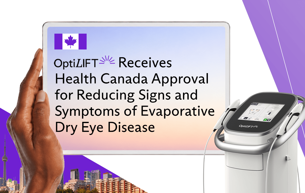
Lumenis OptiLIFT Dynamic Muscle Stimulation Device receives Health Canada Approval for Reducing Signs and Symptoms of Evaporative Dry Eye Disease

The Potential Benefits of Periorbital Transcutaneous Electrical Stimulation for the Management of Dry Eye Disease

Sisters Grow Practice Into Medical Hub for Dry Eye Treatment
Discover new
possibilities
Download OptiLIGHT and OptiPLUS info kit

1. Liu et al. (2017), Am J Ophthalmol 183-190;
2. Yin et al. (2018), Curr Eye Res 43(3):308-313;
3. Kassir et al. (2011), J Cosmet Laser Ther 13(5):216-322;
4. Papageorgiou et al. (2008), Br J Dermatol 159(3):628-632;
5. Prieto et al. (2002), Lasers Surg Med 30(2):82-85;
6. Dell et al. (2017) Clin Ophthalmol 11:817-827;
7. Toyos & Briscoe (2016), J Clin Exp Ophthamol 7:6.
PB-00043680, Rev A
PB-00054400, Rev A